|
The most accurate method for diagnostics of a diamond that contains filled fractures is examining the diamond under magnification. The diagnostics implies the use of a gemological binocular microscope, equipped with a fiber-optic light source, a polarization filter, and accessories for illuminating the studied sample in accordance with the "dark field" technique.
 Features of diamonds treated by fracture filling
 |
- A flash effect is observed in all diamonds treated by fracture filling and can be considered as the most reliable feature of a diamond treated in such a way. The flash effect is an iridescent flash of light, which occurs at the boundaries of either a filled fracture or a laser-drilled hole when the studied sample is slowly rotated under illumination.
In the case when the viewed sample is illuminated in accordance with the "dark field" technique, secondary reflection of light may occur at the diamond facets. The secondarily reflected light can form some bright-field illuminated regions.
Under the "dark field" illumination, the flash-effect colors are usually yellowish orange, violet-purple, and pink. Rarely, yellow, blue, green, and red colors can be observed.
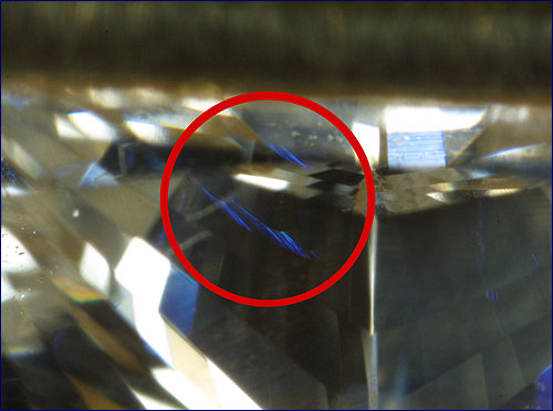
Under the "bright field" illumination, the flash-effect colors are usually blue, blue-green, green, and yellow. More rarely, violet can be observed.
| Blue flash effect in a natural fracture filled polished diamond
|
|
 |
The following hints about observing the flash effect can help one to find this effect in the sample under study:
- a filled fracture may be characterized by different flash-effect colors in different regions of the diamond (the viewing angle being the same);
- when rotating the stone, one portion of the fracture may change the flash-effect colors, while another portion of the fracture maintains its color;
- a flash-effect flare may multiply reflect at the diamond facets; therefore, it is often easier to notice these reflections than to locate the filled fracture.
Interference of light, which occur in thin portions of some non-filled fractures and looks like alternating rainbow-colored fringes, can be easily confused with the flash effect. The following hints about observing the interference and the flash effect make it possible to distinguish between these two phenomena:
- the interference coloring of a non-filled fracture are best observable when the viewing direction slightly differs from the perpendicular to the fracture plane, while the flash effect is strongest for the viewing direction almost perpendicular to the plane of a filled fracture;
- non-filled fractures often have a "feather-like" structure; such a structure is never observed for filled fractures;
- should the observer place a polarization filter between the studied diamond and himself, rotation of the filter will shift the colored interference fringes, while the flash-effect colors just become brighter or darker.
In near-surface fractures or on a diamond surface, orange-brown spots induced by natural iron compounds or green spots induced by natural radiation can be sometimes observed. Such spots can be confused with the flash effect. Unlike the flash effect, these spots are observable for a wider range of viewing angles. This feature enables distinguishing between the spots of natural coloration and the flash effect.
In some cases, the flash-effect colors become much more noticeable when the studied diamond is illuminated with a fiber-optic light source.
- Flow structure. In filled fractures, the filling material may have a glassy structure (there are striaes there). Such a structure is never observed in non-filled fractures.
- Captured bubbles are regions of incomplete filling of a fracture. They can be relatively large and flattened or small and grouped. In the latter case, the captured bubbles form "fingerprint" patterns. When a diamond is viewed under "dark field" illumination, the captured bubbles strongly reflect light and look brighter than the rest of the stone.
- Regions of incomplete near-surface filling are shallow near-surface defects formed as a result of partial removal of a filling material, diamond cleaning, or other processes. These defects look like white fringes or ribbons when viewed under "dark field" illumination.
- Fractured structure of a filling material looks like a web and appears when the filling material cracks. This diagnostic feature is relatively rear for diamonds and is associated with relatively thick fractures. Such a structure can be found in a material filling a laser-drilled hole.
- Filling material turbidity regions are cloud-like portions of the filling material, which have partially lost their transparency.
- Filling material color can be noticed in relatively thick fractures, both filled and laser-drilled. The color is usually light brown, brownish yellow, or orange-yellow.
|
|
Recommended procedure for diagnostics of diamonds treated by fracture filling |
- Carefully clean the sample under study before starting the diagnostics.
- Examine the sample under a gemological binocular microscope. Attentively search for features of diamonds treated by fracture filling.
- To search for the main diagnostic feature of a treated diamond, known as the flash effect, slowly swing the sample since this effect is noticeable only for certain viewing angles. Attentively examine all zones of the stone, one after another, to make the diagnostics reliable
|
|