|
Inclusions are main diagnostic features determined under magnification. To examine the inclusions, a binocular microscope with a magnification factor of 40x and higher is used.
When working with such a microscope, the following illumination modes are recommended:
- dark-field illumination or backlighting;
- fiber-optic lighting - to study inclusions in more detail.
It is also recommended to use the following ways of gripping the stone in the stoneholder of the microscope:
- by girdle;
- by table and culet;
- other ways, depending on the cut style of the sample under study.
Diamond inclusions can be tentatively divided into four groups:
- those inclusions whose presence is a direct diagnostic feature of the synthetic origin of the studied diamond;
- those inclusions whose presence is an indirect diagnostic feature of the synthetic origin of the studied diamond;
- those inclusions whose presence is a direct diagnostic feature of the natural origin of the studied diamond;
- those inclusions whose presence is an indirect diagnostic feature of the natural origin of the studied diamond.
In some cases, to precisely identify the inclusions (for example, to determine their chemical composition), more complex laboratory methods of material characterization, such as Raman spectroscopy or microprobe analysis, are used.
 Inclusions that are direct diagnostic features of synthetic diamonds
 |
- Tenite, Fe0,3Ni0,7. Tenite inclusions are most common for synthetic diamonds. The size of tenite crystals may amount to 1-3 mm. The shape of the crystals is the following: dots, elongated and flattened inclusions (often oriented along the growth planes of the diamond), or phantoms. These inclusions may be framed in cut negative crystals. Most often, they are located near the nucleus of a diamond crystal. Flattened tenite inclusions are usually located in the center of a growth sector of an octahedron or a cube. They glitter like a metal when viewed in reflected light. In transmitted light, they are opaque. Tenite crystals are magnetic.
- Wustite, Fe0,95Ni0,05O. These are flattened triangular crystals with {111} facets, belonging to 2D or 3D dendrites. The crystals are opaque and glitter like a metal. Their size may amount to 0.2 mm.
- Spinel (of various composition with prevalence of Fe). These are needle-like inclusions grouped into fan-like bunches with the maximum length of 2 mm. Spinel crystals are black (dark). They may be located in all growth pyramids, and are subnormally elongated with respect to the facets of the original diamond crystal.
- Cavities filled with negative diamond crystals.
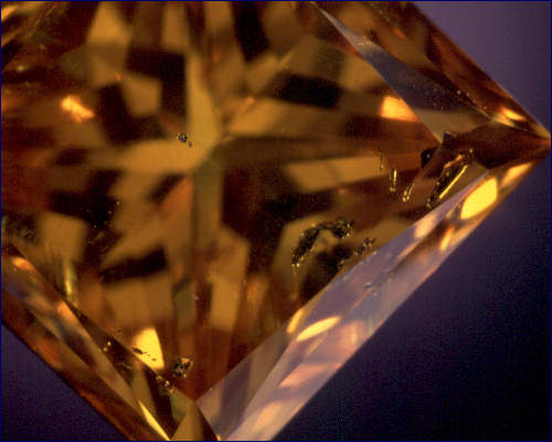
| Inclusion of metallic flux in a synthetic polished diamond
|
|
 |
|
|
Inclusions that are indirect diagnostic features of synthetic diamonds |
- Dispersed inclusions of various composition, looking like clusters of dots, "clouds", or "torches".
- Diamond in diamond. These are irregularly distributed colorless low-profile crystals whose size may amount to 0.2 mm. The most common shape of these crystals is octahedral and cuboctahedral.
|
|
Inclusions that are direct diagnostic features of natural diamonds: |
- Garnet (pyrope). These are isometric (sometimes, slightly elongated) grain-like inclusions whose surface is formed by induction facets. Small pyrope inclusions look like bright dots, those with a size of about 0.1 mm look like bubbles, while larger inclusions are pink or red colored. Pyrope crystals are transparent when viewed in transmitted light.
- Olivine. These are transparent, slightly or moderately elongated crystals. Small and medium-size olivine inclusions look bright (colorless), while larger ones have light-green or green coloration.
- Diopside (chrome diopside). These crystals are similar to olivine and differ from it by a more elongated shape and stronger coloration.
- Sulfides. These mineral inclusions are most common for natural diamonds. They are often called "graphite" or "coal", which is incorrect. The inclusions are dark (black) and opaque, and glitter in a non-metallic manner. The sulfide inclusions look like dark dots (separate or grouped), "cakes", "rosettes", or elongated foreign bodies. Sulfide inclusions are sometimes surrounded by fractures. Often, fractures start from inclusions. In this case, a fracture portion adjacent to an inclusion may look dark.
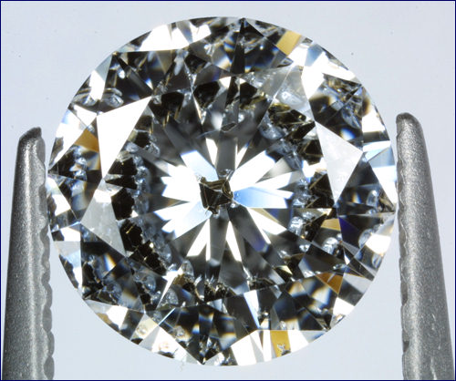
| Red garnet (pyrope) included in a natural polished diamond
|
|
 |
| Red garnet (pyrope) included in a natural polished diamond
|
|
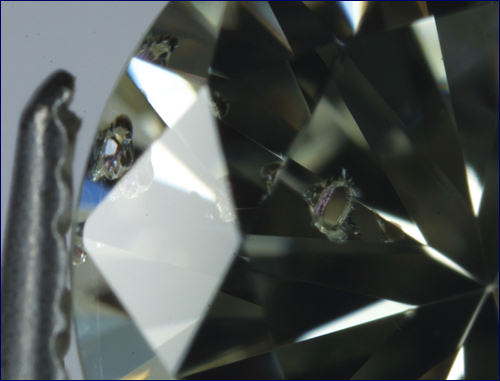 |
| Inclusion of spinel crystal in a natural polished diamond
|
|
 |
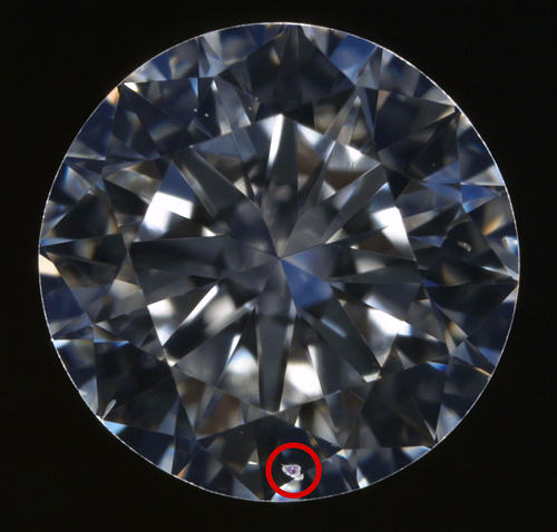
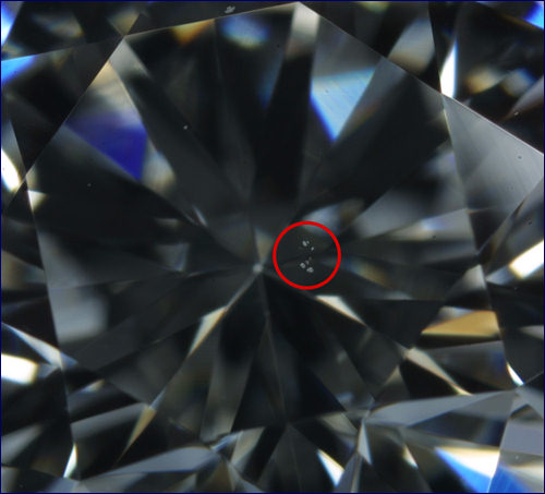
| Group of white pinpoints in a natural polished diamond
|
|
 |
|
|
Inclusions that are indirect diagnostic features of natural diamonds |
- Ilmenite. These are dark needle-like inclusions, usually solitary.
- Spinel (chromospinelide). These are isometric inclusions whose shape is similar to that of sulfide inclusions.
- Diamond in diamond.
|
|
Recommended procedure for studying inclusions: |
- Carefully clean the sample under study before starting the tests.
- Take a microscope and switch on either backlight or "dark field" illumination.
- Thoroughly examine the sample under the microscope.
- During the examination, pay attention to the presence of inclusions and their properties (color, luster, relief, etc.).
- To observe the stone in more detail, use a fiber-optic light source at larger magnification.
- Identify the observed inclusions using the description of inclusions that occur in natural and synthetic diamonds.
|
|